

dental x ray positioning guide
Master dental X-ray positioning with our comprehensive guide. Learn proper techniques for clear images and accurate diagnoses.
Dental x-ray positioning is fundamental for accurate diagnostic imaging, ensuring clear visuals of tooth structures and surrounding tissues․ Proper techniques minimize retakes and radiation exposure, enhancing patient safety and diagnostic efficiency․
Overview of Dental X-Ray Positioning
Dental x-ray positioning involves precise techniques to capture accurate images of dental structures․ Proper alignment of the x-ray beam, patient positioning, and film placement are critical․ This process ensures clear visualization of teeth, bones, and surrounding tissues, aiding in diagnoses․ Positioning guides, such as the iM3 Dental X-Ray Positioning Kit, help standardize procedures, reducing retakes and radiation exposure․ Effective communication with patients enhances cooperation, while proper techniques minimize discomfort and ensure safety․ Regular updates in methods and tools optimize results, making dental radiography more efficient and reliable for both clinicians and patients․
Importance of Proper Positioning in Dental Radiography
Proper positioning in dental radiography ensures clear visuals of tooth structures and surrounding tissues, minimizing retakes and radiation exposure․ Accurate alignment enhances diagnostic precision, allowing early detection of issues like decay or fractures․ Misalignment can obscure critical details, leading to missed diagnoses or unnecessary treatments․ Correct positioning also reduces patient discomfort and streamlines the process, improving overall efficiency․ By adhering to established techniques, dental professionals can deliver high-quality care while maintaining patient safety and confidence․ Proper positioning is thus a cornerstone of effective and ethical dental radiography practice․
Types of Dental X-Rays
Dental x-rays are categorized into intraoral and extraoral types․ Intraoral x-rays include periapical and bitewing views, while extraoral x-rays include panoramic and cephalometric radiographs․ Understanding these types is essential for accurate imaging and diagnostics․
Intraoral X-Rays
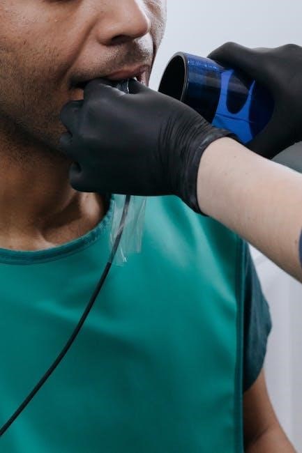
Intraoral x-rays are taken inside the mouth, providing detailed images of individual teeth and surrounding bone․ Common types include periapical, bitewing, and occlusal views․ Bitewing x-rays detect interproximal caries and bone loss, while periapical images show entire teeth and roots․ Proper positioning involves placing the x-ray sensor or film parallel to the dental arch, ensuring accurate alignment․ This technique minimizes distortion, allowing for precise diagnosis of decay, fractures, or periodontal issues․ Clear communication with the patient is key to achieving correct positioning and ensuring comfort during the procedure․ Intraoral x-rays are essential for comprehensive oral health assessment․
Extraoral X-Rays
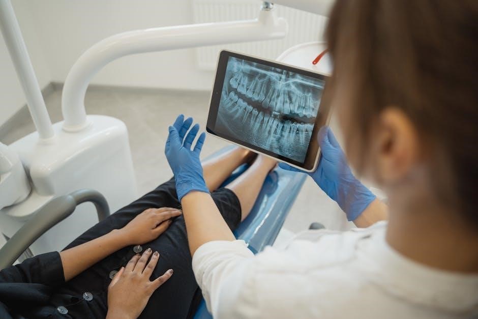
Extraoral x-rays capture images of the teeth and jaw from outside the mouth, offering a broader view of the dental structure․ They are ideal for diagnosing issues like impacted teeth, fractures, or pathologies․ Techniques include panoramic and cephalometric x-rays, which provide comprehensive overviews․ These x-rays are particularly useful for patients with limited mouth opening or when assessing large-scale abnormalities․ Proper positioning involves aligning the patient correctly with the x-ray beam to ensure accurate images․ Extraoral x-rays reduce retakes and discomfort, making them a valuable tool in dental diagnostics․ Their versatility enhances overall patient care and treatment planning․

Preparation for Dental X-Ray Procedures
Effective preparation involves clear patient communication, explaining the procedure to reduce anxiety, and ensuring the patient understands the importance of remaining still during imaging․
Patient Preparation and Communication
Effective patient preparation and communication are crucial for successful dental x-ray procedures․ Start by explaining the process clearly, addressing any concerns or anxieties the patient may have․ Use visual aids to demonstrate positioning and ensure the patient understands the importance of remaining still during the x-ray․ Provide instructions in a calm and reassuring manner to reduce stress․ Ensure the patient is comfortable and properly positioned to avoid movement and ensure accurate images․ Always obtain informed consent and answer questions to build trust and cooperation․ Clear communication enhances patient compliance and improves the overall outcome of the procedure․
Positioning the Patient Correctly
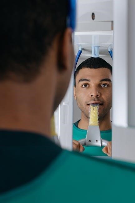
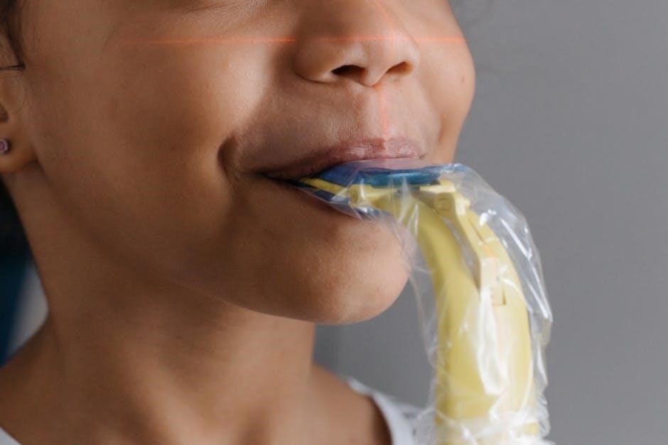
Correct patient positioning is essential for obtaining accurate and diagnostic-quality dental X-rays․ The patient should be seated upright with their head stabilized to ensure alignment of the X-ray beam; For intraoral X-rays, the patient’s mouth must be open wide enough to accommodate the sensor or film, with the tongue resting gently behind the upper teeth; Proper angulation of the X-ray beam ensures that the desired tooth structures are captured without distortion․ Clear communication and guidance are crucial to help the patient maintain the correct position, minimizing retakes and ensuring safety․ Using positioning aids like bite blocks or headrests can enhance stability and accuracy․
Dental X-Ray Positioning Techniques
Modern techniques involve precise patient alignment, using tools like the iM3 X-Ray Positioning Kit, to ensure accurate imaging and minimize retakes, focusing on clear tooth visualization․
Intraoral X-Ray Positioning Methods
Intraoral X-rays are taken inside the mouth, providing detailed images of individual teeth and surrounding structures․ Common methods include the paralleling technique, where the X-ray beam is perpendicular to the dental film and parallel to the tooth long axis, minimizing distortion․ The bisecting angle technique involves angling the beam to bisect the angle between the tooth and the film, useful for posterior teeth․ Proper positioning ensures accurate diagnostics, reduces retakes, and optimizes radiation safety․ Tools like positioning kits and dental charts aid in precise alignment, ensuring clear visuals of tooth structures for effective treatment planning․
Extraoral X-Ray Positioning Techniques
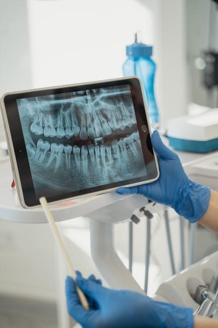
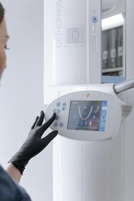
Extraoral x-ray positioning involves placing the film or sensor outside the mouth, offering a broader view of dental and facial structures․ Techniques like panoramic, cephalometric, and CBCT (Cone Beam Computed Tomography) require precise patient positioning․ For panoramic X-rays, the patient stands upright with the head centered and bite closed․ Cephalometric X-rays need the head aligned in a specific plane for accurate skull measurements․ Proper alignment and angulation are critical to avoid distortion and ensure diagnostic accuracy․ These techniques are essential for evaluating complex cases, such as impacted teeth or orthodontic planning, and provide a comprehensive view of the jaw and surrounding anatomy․
Common Errors and Mistakes to Avoid
Common errors include improper patient positioning, insufficient anatomical knowledge, and inadequate communication, leading to retakes and diagnostic inaccuracies․ Proper training and clear patient instructions are essential to avoid these mistakes․
Identifying and Correcting Positioning Errors
Positioning errors can lead to unclear images, retakes, and increased radiation exposure․ Common mistakes include improper alignment of the X-ray beam, incorrect angulation, and inadequate patient preparation․ To correct these, ensure the beam is perpendicular to the dental film or sensor․ Adjust the patient’s head position to achieve proper alignment․ Use positioning guides or stickers for consistency․ Communicate clearly with patients to minimize movement․ Regular training and equipment calibration can prevent such errors, ensuring accurate and safe radiographic outcomes․ Correcting these issues enhances diagnostic quality and patient safety․ Proper techniques must be prioritized in every procedure․

Safety Measures in Dental Radiography
Protective gear like lead aprons and thyroid collars is essential to minimize radiation exposure․ Strict adherence to safety guidelines ensures patient and operator protection during procedures․
Radiation Protection and Safety Guidelines
Minimizing radiation exposure is crucial for patient safety and adhering to regulatory standards․ Dental professionals must use lead aprons, thyroid collars, and digital sensors to protect patients․ Strict adherence to the ALARA principle ensures doses are as low as reasonably achievable․ Regular equipment calibration and proper positioning techniques reduce unnecessary exposure․ Patients with high risk factors or pregnancy should receive extra precautions․ Staff should monitor radiation levels and undergo training to maintain safety protocols․ These guidelines ensure a balance between diagnostic quality and patient protection, promoting a safe environment for both patients and practitioners․
Interpreting Dental X-Ray Images
Accurate interpretation requires a solid foundation in dental anatomy and pathology, enabling recognition of normal structures and identification of abnormalities like decay, fractures, or bone loss․
Basic Interpretation Techniques
Interpreting dental x-rays requires a thorough understanding of dental anatomy and pathology․ Start by evaluating the clarity and positioning of the image․ Identify normal structures like enamel, dentin, and pulp․ Look for abnormalities such as caries, fractures, or signs of infection․ Use a systematic approach to examine each tooth and surrounding tissue․ Compare affected areas with adjacent or opposing teeth for contrast․ Note any unusual radiopacity or radiolucency․ Correlate findings with clinical symptoms to confirm diagnoses․ Proper interpretation ensures accurate detection and treatment planning, making it a critical skill in dental care․
Choosing the Right Equipment
Selection Criteria for Dental X-Ray Equipment
Selecting the right dental x-ray equipment ensures precise imaging and patient safety․ Consider image quality, ease of use, and compliance with radiation safety standards․ The iM3 Complete X-Ray Positioning Kit is a recommended tool for accurate positioning․
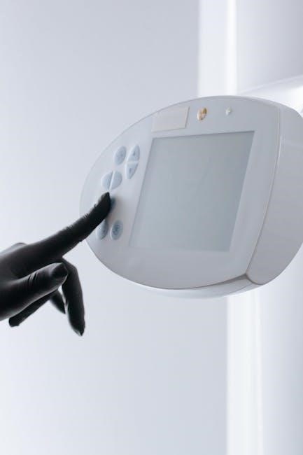
Selecting the right dental x-ray equipment is crucial for producing high-quality images while ensuring patient safety․ Key factors include image resolution, ease of positioning, and radiation dose control․ Durability and compatibility with digital systems are also essential․ Consider the space and budget constraints of your practice․ Modern equipment often features advanced safety measures, such as timers and collimators, to minimize radiation exposure․ Additionally, look for equipment with user-friendly interfaces and customizable settings to enhance workflow efficiency․ Investing in a positioning kit, like the iM3 Complete X-Ray Positioning Kit, can simplify the process and improve accuracy․ Prioritize equipment that balances performance, cost, and patient comfort for optimal outcomes․
Troubleshooting Common Issues
Resolving Technical Difficulties in X-Ray Positioning
Common issues include misalignment, patient movement, or equipment malfunctions․ Using tools like the iM3 Positioning Kit and referencing guides can help resolve these challenges effectively․
Technical difficulties in x-ray positioning often arise from incorrect alignment or device malfunctions․ Start by identifying the source of the issue, such as misangled beams or improper patient positioning․ Use tools like the iM3 X-Ray Positioning Kit for precise alignment․ Adjust the patient’s posture and ensure the x-ray beam is parallel to the dental film․ Retake images if necessary, and refer to guides like PS011 on Youtube for troubleshooting tips․ Regular equipment maintenance and staff training can prevent recurring issues, ensuring high-quality radiographs and efficient diagnostic processes․
Mastering dental x-ray positioning is essential for producing high-quality radiographs that aid in accurate diagnoses․ Proper techniques ensure patient safety, reduce radiation exposure, and enhance image clarity․ By following established guidelines and continuously refining skills, dental professionals can optimize outcomes․ Regular training and adherence to safety protocols are vital for maintaining excellence in dental radiography․ This guide provides a comprehensive framework to help practitioners achieve precise and reliable results, ultimately improving patient care and diagnostic confidence․